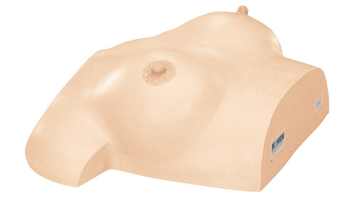
Inspection and Palpation of Breast Cancer Training Model (Precision Type)
The incidence of breast cancer is high in women in the prime of their life.
Should breast cancer develop, cancer cells can readily enter the bloodstream and spread throughout the whole body.
If detected and treated at an early stage while still localized, the chances of complete recovery are very high.
Since the breast is an organ located close to the body surface, any change that develops on the skin can be seen and changes in the breast can be palpated.
Based on these characteristics, we developed this model so that changes in the skin that appear with breast cancer can be understood and knowledge of breast cancer symptoms can be acquired through visual inspection.
Most breast cancers are detected by palpation of a "lump".
The smaller the lump, the more readily it heals after treatment. From this perspective, careful self-examination every month is recommended in addition to professional examination by a doctor. If a lump or mass is detected, a specialist should immediately be consulted.
This model is a teaching aid for detecting breast cancer symptoms and for training women in self-examination.
Features
The size and texture of the model is made to be as realistic as possible. Pathological symptoms such as lumps and skin changes may be slightly exaggerated in the model, but the conditions and feel of the lesions are created with the utmost accuracy. This model offers an ideal educational tool for medical students, nursing students and health nurses.
The model can be effectively used as a teaching tool for self-examination of breast cancer by the general public through mass screening and for training in detection of breast cancer (intermittent breast cancer) outside the doctor's office.
Symptoms of Breast Cancer
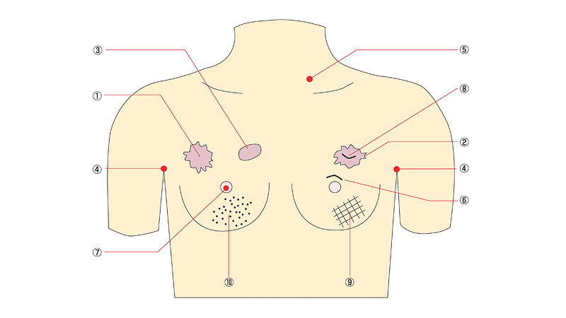
Symptom | Number | Description |
| Lumps | 1. | Hard lumps. Cancer is suspected due to the irregular surface. |
| 2. | ||
| 3. | Soft lump. Cancer is suspected after palpation | |
| Lymph node metastasis | 4. | Lymph nodes in the axillary region (armpits) |
| 5. | Hard lymph node in the neck | |
| Nipple changes | 6. | Displacement or depression of the nipple |
| 7. | Eczematous change (sore) [Paget's cancer] | |
| Skin changes | 8. | Skin dimpling |
| 9. | Partial edema of the skin, “orange-peel" appearance | |
| 10. | Skin redness and swelling (inflammatory breast cancer) |
Specifications
| Main body | Approx.35(L) × 43(W) ×18(H)cm | Approx.3.7kg |
| Storage Case | Approx.42(L) × 47(W) ×22(H)cm | Approx.3.5kg |


