Introduction to Paramecium

AUSTRALIAN CURRICULUM ALIGNMENT
- The theory of evolution by natural selection explains the diversity of living things and is supported by a range of scientific evidence
- Describing biodiversity as a function of evolution
BACKGROUND:
Paramecium are ciliated Protists, and are surprisingly complex for such tiny organisms. Paramecium have previously been classified under Domain Eukarya; Kingdom Protista; and Phylum Ciliophora. Currently, ciliates are grouped with dinoflagellates and sporozoans in the clade Alveolates.In this practical, students are tasked with observing a Paramecium culture under the microscope. Students are tasked with identifying key features of Paramecium and observing their movement. This practical is a great introduction to microscopic life forms and provides an opportunity for students to practice sampling and wet mounting. Students may work individually or in pairs.
PREPARATION - BY LAB TECHNICIAN
Preparing the Culture
- As soon as your culture of Paramecium arrives, loosen the lid on the container and aerate the culture using a plastic pipette.
- To aerate the water, hold the pipette tip into the culture water and squeeze the bulb. Release the bulb and allow it to fill with air once again. Repeat this step 4 more times to assist in replacing the oxygen depleted during shipping.
- Replace the lid loosely, but do not screw too tightly.
- Store in a dark cupboard away from direct sunlight.
Preparing Workstations
- Provide each workstation with the following materials.
- Paramecium Culture
- Protoslo Solution
- Microscope Slides
- Transfer Pipettes
- Coverslips
METHOD - STUDENT PRACTICAL
-
Collect the Paramecium culture and place it on your workstation.
- Observe the culture with the naked eye and note what you see.
- To remove a sample from the culture, compress the bulb of the pipette between your thumb and forefinger and lower the tip to the bottom of the culture jar. Then, gently release the pressure on the bulb and allow the sample to be sucked into the pipette. We recommend removing the sample from the bottom of the jar as the Paramecium will be in the greatest concentration there.
- Deliver 2–3 drops of your sample onto a microscope slide along with a drop of Protoslo. Mix thoroughly and carefully place a coverslip on the top.
- To locate the Paramecia on the slide, observe under the low-power objective of your microscope and centre the Paramecium in the microscopic field. Once you have located it, rotate to the high-power objective and refocus to observe the paramecium in greater detail.
OBSERVATION AND RESULTS
Ask students to study the paramecium under the microscope and observe:-
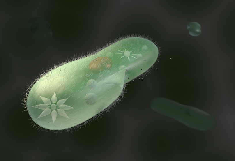
Oral groove: Appearing as a depression on one side of the body. The oral groove extends from the anterior end to beyond the middle. The oral groove serves to guide food particles, primarily bacteria, into the pharynx (gullet). Food particles collect in the bottom of the cavity and are budded off into new food vacuoles.
- Food vacuoles: Found in the cytoplasm, the food vacuoles are spherical structures of varying sizes that store food during digestion.
- Contractile vacuole: Frequently expanding to a larger size and collapsing suddenly, the contractile vacuole is a clear spherical structure. It contracts by discharging its contents into the surrounding medium and is external to the cell itself. Students should observe the number of contractile vacuoles that are observable in the Paramecium.
- Radiating canals: Connected to the contractile vacuoles, these channels in the cytoplasm frequently often give a star pattern to the contractile vacuoles.
- Cilia: Cilia are hair-like organelles that span the surface of the paramecium and enable locomotion. The cilia also function for food retrieval, cilia in the oral groove direct particles towards the oral groove into the pharynx in a sweeping motion.
- As students are observing the Paramecium swim, ask them to note whether there are any indications the organism has an anterior end. Suggest students switch to low power during this section of the observation.
- Request students observe the pattern of movement of the paramecium as it swims and draw a diagram that illustrates what they see. Students can draw a diagram of what occurs when the Paramecium encounters an obstacle.
- To improve memory retention of Paramecium physiology, ask students to draw a Paramecium and label all the parts they observed in this practical.
EXTENSION EXERCISE:
-
An exciting way to see the Paramecium digestive system functioning is to feed the paramecium a yeast/congo red solution. As the food moves is digested by the Paramecium pH changes will cause the Congo Red to change from red to blue.
- Paramecium are a ciliated Protist that have previously been classified under Domain Eukarya, Kingdom Protista, and Phylum Ciliophora. Currently ciliates are often grouped with dinoflagellates and sporozoans in the clade Alveolates. The term protist, is currently used to describe a grade of organisation that includes unicellular (or multicellular without tissues) organisms; such as eukaryotic organisms. It is often referred to as the ‘junk drawer’ kingdom. To explore classification of microorganisms, you may like to discuss why the Kingdom Protista is often referred to as the ‘junk drawer’ kingdom.
TEACHER TIP:
- While the students are undertaking the practical, you may like to take the opportunity to discuss avoidance behaviour. If a Paramecia encounters negative stimulus, it can rotate up to 360 degrees in order to escape. Ask students to consider how this trait may have evolved.
 Time Requirements
- 45 min
 Material List
 Complimentary products
![]() Safety Requirements
Safety Requirements
-
Wear appropriate personal protective equipment (PPE).
- Wash your hands thoroughly before and after the practical.
- Do not release any organisms into the environment.
- Disinfect work areas before and after the practical.
